Sure! Here’s a fresh version of the science story that’s engaging and understandable:
—
In the fascinating world of embryonic development, a recent breakthrough shines a light on how tiny stem cells journey to form essential layers of life. Thanks to innovative research from the University of Michigan, we’re now one step closer to understanding the early stages of human development, with potential implications for preventing birth defects and improving reproductive health.

A recent study led by Ph.D. candidate Bohan Chen and supervised by Idse Heemskerk at the University of Michigan Medical School highlights enhancements to a popular scientific model known as gastruloids. These miniature models replicate the early stages of embryo formation, a process called gastrulation, without the ethical concerns that come with studying actual embryos.
Gastruloids are created from stem cells and are a significant step in simulating the journey of embryonic cells. During gastrulation, cells differentiate into three key layers: the ectoderm (which forms the skin and nervous system), the mesoderm (which becomes muscles, bones, and the heart), and the endoderm (which develops into vital organs like the liver and lungs).
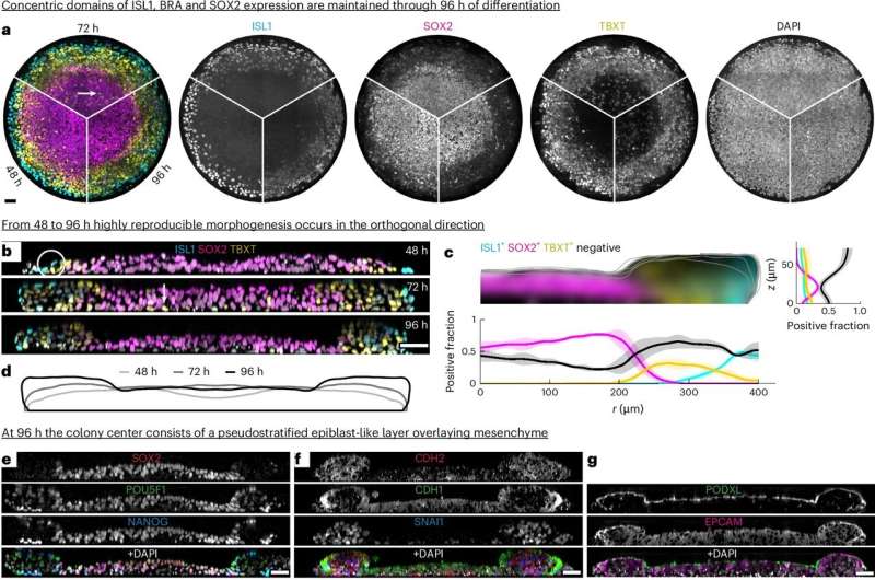
Typically, gastruloids would only grow for about two days before becoming disorganized. However, the research team made notable improvements, experimenting with different growth media and conditions. To their delight, the gastruloids not only survived beyond those two days but began to show intriguing developments.
The breakthrough moment came when researchers observed the mesoderm cells migrating beneath the initial layer, forming a multi-layered structure much like that in a real embryo. This migration is so pivotal, yet its mechanisms remain largely mysterious. Heemskerk noted that this enhanced model gives us the chance to explore the signals that guide these migrating cells—a crucial step in understanding congenital conditions like heart defects.
Even more fascinating was the discovery that these mesoderm cells exist in different subtypes, each expressing unique genes. By observing these gene expressions, scientists can predict the potential fate of each cell, illuminating the age-old mystery of whether cells know their destiny before they even begin to move.
Heemskerk expressed a sense of excitement for future investigations using their improved model, stating, “Not only does this simple system help us observe phenomena that are hard to replicate in more complex conditions, but it also allows for ethical research while uncovering deep biological truths.” This optimistic outlook hints at a promising future in the quest to better understand human development and prevent birth-related complications.
More information:
Bohan Chen et al, Extended culture of 2D gastruloids to model human mesoderm development, Nature Methods (2025). DOI: 10.1038/s41592-025-02669-4
If you would like to see similar science posts like this, click here & share this article with your friends!


